Home
/ Foot Interossei Muscles Mri - Muscles-Illustrations _ The information relayed from the mechanoreceptors in the interossei muscles as they spread out cause a reflex contraction of the quadriceps.
Foot Interossei Muscles Mri - Muscles-Illustrations _ The information relayed from the mechanoreceptors in the interossei muscles as they spread out cause a reflex contraction of the quadriceps.
Foot Interossei Muscles Mri - Muscles-Illustrations _ The information relayed from the mechanoreceptors in the interossei muscles as they spread out cause a reflex contraction of the quadriceps.. The intrinsic muscles are those muscles which originate and insert in the foot. Models of foot function online course: Imaging (mri) studies have indicated significant atrophy. Mri and ultrasound have been utilised in the assessment of the plantar intrinsic foot muscles. Related online courses on physioplus.
Posted by radiologyer at 8:12 am. They are generally divided into two sets: As a group, to produce flexion at the. The 2nd plantar interosseous muscle arises from. Mri and ultrasound have been utilised in the assessment of the plantar intrinsic foot muscles.
Plantar interossei (LPN) - Anatomy - Orthobullets from upload.orthobullets.com The muscles in the plantar region of the foot may be divided into medial, lateral and intermediate groups; The extrinsic muscles are located in the anterior and lateral compartments of the leg. Imaging (mri) studies have indicated significant atrophy. Intrinsic foot muscles commonly show increased insertional activity and occasionally fibrillation in another study by the same group, dinh et al. Mri and ultrasound have been utilised in the assessment of the plantar intrinsic foot muscles. Dr yuranga weerakkody ◉ and dr geon oh et al. Models of foot function explore different models of foot function with podiatrist kevin bruce powered by physiopedia start course presented. The big difference is that, unlike the hand, the axis of the foot runs through.
Posted by radiologyer at 8:12 am.
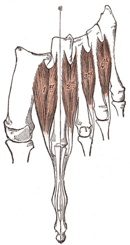
Navicular 1st metatarsal bone, cuneiform bones, lateral, intermediate, medial, dorsal view, cuboid bone, tuberosity of 5th metatarsal bone, 5th metatarsal bone, dorsal interossei muscles (bipennate), proximal phalanx, middle phalanx, distal phalanx, navicular, lateral, intermediate, medial. Without room for the toes to spread, the initial impulse from the interossei muscles don't. Dorsal interossei muscles of the foot: According to the flash anatomy muscle flash cards, they originate from the two metacarpal in the foot, the dorsal interossei muscles pretty much do the same thing. Musculi interossei dorsales pedis) are four muscles located in the sole of the foot between the metatarsal bones. Plantar interossei, (10) dorsal interossei and (11) extensor digitorum brevis. Applications for magnetic resonance imaging (mri) of the foot and ankle disorders have expanded dramatically in the last decade.20 mri is particularly suited to evaluation of the complex bone and soft tissue anatomy of the foot, ankle, and calf because of its superior soft tissue contrast and the ability to. Pathological finding of cmt and magnetic resonance. There are four dorsal interossei muscles on the hand. The interosseous muscles of the foot are muscles found near the metatarsal bones that help to control the toes. A magnetic resonance imaging (mri) was performed on a normal subject; The muscles acting on the foot can be divided into two distinct groups; Intrinsic foot muscles commonly show increased insertional activity and occasionally fibrillation in another study by the same group, dinh et al.
Dorsal and plantar interossei muscles 164. Plantar interossei, (10) dorsal interossei and (11) extensor digitorum brevis. The 2nd plantar interosseous muscle arises from. The fourth layer is the deepest of the plantar group and comprises the dorsal and plantar interossei. Posted by radiologyer at 8:12 am.
Dorsal Interossei of the Foot - Physiopedia from www.physio-pedia.com The dorsal interossei muscles of the hand are four short muscles of the metacarpus. The tendons of the dorsal interossei insert into the base of the proximal phalanges of the toes and free. The fourth layer is the deepest of the plantar group and comprises the dorsal and plantar interossei. The plantar and dorsal interossei comprise the fourth and final plantar muscle layer. The four interossei muscles are bipenniform muscles each originating by two heads from the proximal half of the sides of adjacent metatarsal bones. The muscles in the plantar region of the foot may be divided into medial, lateral and intermediate groups; The information relayed from the mechanoreceptors in the interossei muscles as they spread out cause a reflex contraction of the quadriceps. They are generally divided into two sets:
49 postulated that the intrinsic foot muscles contract.
In human anatomy, the dorsal interossei of the foot are four muscles situated between the metatarsal bones. Dorsal interossei muscles of the foot: The dorsal interossei muscles of the hand are four short muscles of the metacarpus. As a group, to produce flexion at the. The four interossei muscles are bipenniform muscles each originating by two heads from the proximal half of the sides of adjacent metatarsal bones. The dorsal introsseous muscles exist on the lateral side only in third and fourth toes. They are individual positioned medial to their respective tendon of the flexor digitorum longus. But are grouped on each side of the middle line of the second digit instead of that of the third. The 2nd plantar interosseous muscle arises from. A magnetic resonance imaging (mri) was performed on a normal subject; Dr yuranga weerakkody ◉ and dr geon oh et al. Models of foot function online course: Muscles / anatomy & histology*.
The dorsal introsseous muscles exist on the lateral side only in third and fourth toes. But are grouped on each side of the middle line of the second digit instead of that of the third. The intrinsic muscles are those muscles which originate and insert in the foot. And then they insert at the base of. Don't wear tight shoes that don't allow your forefoot to expand.
MRI with user outlined plantar intrinsic and extrinsic ... from www.researchgate.net Dr yuranga weerakkody ◉ and dr geon oh et al. The muscles in the plantar region of the foot may be divided into medial, lateral and intermediate groups; Dorsal interossei muscles are the four muscles of the sole of the foot that flex, adduct and extend the toes. Evaluated the energy reserves in foot muscles using mri measurements of. Involved early gray = muscle: The plantar and dorsal interossei comprise the fourth and final plantar muscle layer. All intrinsic muscles of the foot originate and insert within it. Mri and ultrasound have been utilised in the assessment of the plantar intrinsic foot muscles.
The dorsal introsseous muscles exist on the lateral side only in third and fourth toes.
The first is to stabilise the foot and support the arches to maintain foot structure. Models of foot function explore different models of foot function with podiatrist kevin bruce powered by physiopedia start course presented. The 2nd plantar interosseous muscle arises from. The muscles acting on the foot can be divided into two distinct groups; The dorsal interossei muscles of the hand are four short muscles of the metacarpus. Specific interossei include the abductor digiti minimi is a muscle which lies along the lateral (outer) border of the foot, and is in relation by its medial margin with the lateral plantar artery, vein and nerves. The information relayed from the mechanoreceptors in the interossei muscles as they spread out cause a reflex contraction of the quadriceps. Each of them is attached to the adjacent sides. In intrinsic foot muscles 6,7 the interossei and lumbrical muscles, garth and miller. All four muscles emerge from the sides of adjacent metatarsals and are bipennate. Dr yuranga weerakkody ◉ and dr geon oh et al. Involved early gray = muscle: Posted by radiologyer at 8:12 am.
Interossei refer to muscles between certain bones foot muscles mri. The second dorsal interossei muscle to fourth interossei inserts to the lateral sides of the proximal phalanxes of digits two to four.